
Associate Director (DMPK),
Aragen Life Sciences Private Limited,
Of the 7.9 billion global population in 2021, more than 3 billion people worldwide (over 1 in 3 people) lived with a neurological condition. Neurological disorders are the leading cause of illness & disability worldwide. This article discusses the impact of high pitch loud noise and vibrations on Blood Brain Barrier (BBB) plasticity and manifestation of neurological disease conditions.
The noise standard for mentally stressful tasks is 55 dB. If the noise source is continuous, the threshold level is <55 dB. Accordingly, the sound of >55 dB is considered as noise pollution. The National Institute for Safety & Health (NIOSH) recommends not more than an 8-hour exposure limit of 85 dBA. Per their recommendation, a hearing conservation program should be in place if a worker is exposed to 85 dB x 8-hr work period. Further, if the noise level is 95 dB, the worker can be exposed to the noise for only 4 hrs over a work shift. Extended exposure to noise >90dB triggers stress response. The sound at open air musical concerts peaks >100 dB, which is generally considered unsafe for exposures exceeding 15 min. Exposure to sound at the hearing threshold level for extended periods alters neural activity in auditory processing, emotional and vascular autonomic control regions of the brain. The implosive oscillations caused by acoustic emissions generate shock waves that perforate the weak regions of the BBB near to the lining of the circumventricular organs.
Also, the high pitch sound or vibration causes oxidative stress, which triggers an inflammatory response resulting in loss of BBB integrity. Exposure to sound above 70 dB intensity for long intervals, can cause damage to right frontal cortex histology, cerebral ultrastructure, and BBB, thus allowing blood borne substances to pass across the BBB into the brain tissue. The persistent high pitch noise causes alterations in neurotransmitter release, affects synaptic plasticity, leading to learning and memory impairment.
The mobile phones use wireless technology, with the majority of them operating in a frequency range of about 900 - 1800 MHz, pulse frequency of 217 Hz, pulse width of 577 s, and duty cycle of 12.5%. Only recently has the frequency risen above 2100 MHz.
Mobile phones with multiple transceivers emit more electromagnetic energy than single transceiver versions. Electromagnetic fields from multi-transceiver mobile phones cause oxidative stress in blood and heart tissue of animals and trigger an inflammatory response at BBB. Prolonged exposure to ringtone sound of 72 dB alters the cerebral ultrastructure and causes an increase in sympathetic activity via stress-induced epinephrine increase.
Nitric oxide activity and tumour necrotic factor level increase in the brain tissue after exposure to multi-transceiver mobile phones for extended durations. This relatively selective effect on the cardiovascular and nervous system is because of the high metabolic rate and lipid composition, respectively, which renders them sensitive for oxidative stress response to electromagnetic field exposure during both physiological and pathological processes, such as anxiety, hypertension, and neurodegeneration.
This review discusses the blood brain barrier architecture, function and its disruption by loud sound and phone ring tones or vibrations.
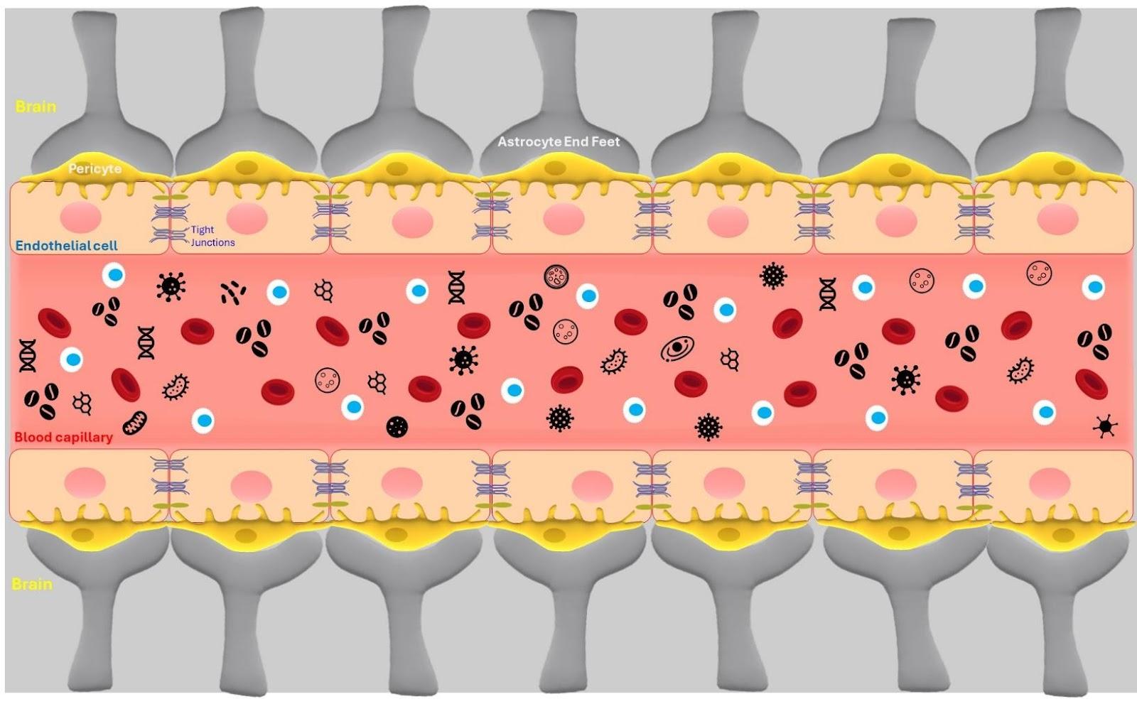
The Blood Brain Barrier
The BBB barrier is a selectively permeable membrane that separates the bloodstream from the brain and flush the metabolites and ions from the brain tissue to blood. Active efflux pumps out the brain materials by ATP binding cassette (ABC) transporters for example P-glycoprotein and Breast Cancer Resistance Protein. A crucial interface between the vascular system and the brain, the BBB acts as a filter to protect neurons from pathogens and inflammation. While it guards the neural tissue from exposure to xenobiotics, it also poses hindrance to treat neurological diseases by limiting the permeability of drugs, and thus achieving therapeutically effective concentration in the affected brain tissue/region. The BBB is composed of endothelial cells (ECs), pericytes and astrocytes.
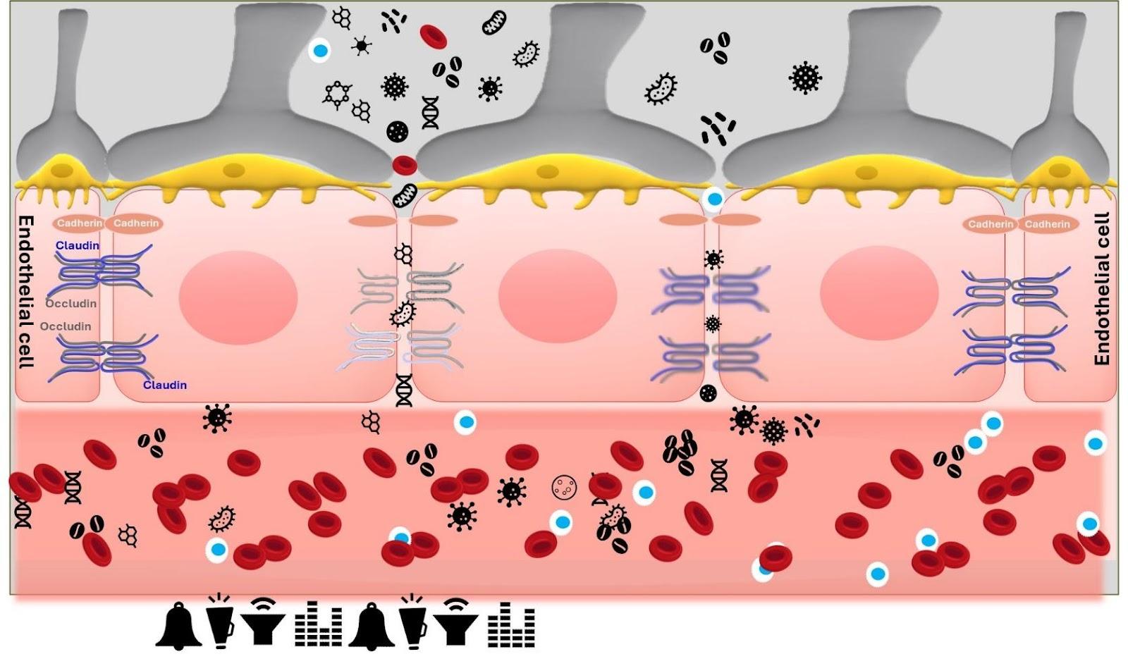
ECs form the walls of the blood vessels and impart mechanical stability. They are connected by tight junctions (TJs) and adherens junctions (AJs).
TJs, the primary regulators of BBB permeability, are formed by three types of transmembrane proteins i.e. 60-kDa integral membrane protein “Occludin”, 20–24/27 kDa “Claudins”, and Junctional Adhesion Molecules (JAMs). Occluding regulated permeability of TJs and Claudin link the membranes of adjacent cells. TJs are anchored to the actin cytoskeleton via cytoplasmic scaffolding protein Zonula Occludens-1, 2 and 3 and heterotrimeric G-proteins.
The high trans-endothelial electrical resistance (TEER) is due to limited paracellular transport governed by tight junctions (TJs). TJs limit passive diffusion through the paracellular space, primarily allowing the lipophilic molecules with specific characteristics and molecular weight cutoff of ≤450 Da to pass through.
By interacting with the cytoskeleton, AJs link ECs to form continuous sheets. AJs are formed by the homophilic interactions between transmembrane cadherin and intracellular catenin proteins to anchor them to actin filaments and tubulin microtubules. Cadherins and catenins mediate pericyte interactions and maintain barrier integrity. Cadherin-10 is predominantly expressed in brain microvessels with BBB phenotypes, while VE cadherin is more abundant in larger pial vessels and in relatively leaky barriers. During acoustic stimulation, the bonds between TJ and AJ proteins are disrupted and decoupled, resulting in transient and reversible opening of the BBB.
To facilitate blood-parenchyma exchange, ECs express receptors and transporters. The large molecules are transported through endocytosis, encapsulating them in caveolin-lined vesicles. This can be specific i.e., receptor-mediated (RMT) or non-specific i.e., adsorption-mediated (AMT) transcytosis. Two common proteins involved in RMT are LDL-receptor related protein-1 and the receptor for advanced glycation end-products (RAGE).
Partially enveloping ECs, the pericytes stay embedded within the vascular basement membrane and regulate the EC development and physiology through intercellular signalling mechanisms. Pericytes extend long membrane processes across the abluminal side of capillaries and cover about 22–37% of the EC surface. Pericytes regulate angiogenesis, infiltration of immune cells and deposit extracellular matrix components.
Astrocytes further encapsulate blood vessels in the brain, extending their end-feet over 99% of the endothelial surface. Astrocytes secrete their own parenchymal basement membrane “glia limitans” in the perivascular space. Astrocytes regulate electrochemical activity, the innate immunity and balance parenchymal water and metabolites.
Transient disruption of Blood Brain Barrier
Under normal circumstances, a small number of mononuclear leukocytes, monocytes and macrophages may enter the CNS via diapedesis. This low intensity leukocyte trafficking across the BBB is for immune surveillance and to exert response to brain infection.
The barrier function, however, is not always rigid and BBB undergoes modulation and regulation, both physiologically and pathologically. The barrier disruption can range from mild and transient TJ opening to chronic barrier breakdown.
Pericytes maintain the integrity of the B-B and their depletion increases BBB permeability via upregulation of endothelial transcytosis. Astrocytes promote BBB formation and integrity via the Hedgehog signalling pathway.
Destabilization of BBB increases the risk of stroke and neurodegenerative diseases, for example vascular dementia. A multitude of factors can disrupt the BBB, which include secreted elements to immune cells and pathogens, reactive oxygen species (ROS), activation of MMPs, and chronic up-regulation of angiogenic factors and pro-inflammatory cytokines. Immunoglobulin G (IgG) extravasation is commonly used as an index of BBB disruption.
Augmentation of paracellular transport relies on weakening the TJ/AJ and allowing inter EC passage. Enhanced paracellular permeation can be achieved by chemical or physical mechanisms. Vasoactive chemical compounds such as histamine, bradykinin, alkylglycerols, tumour necrosis factor, or interferon-γ activate signalling pathways within ECs and increase BBB permeability. Alternative approaches include infusing hyperosmolar agents such as mannitol to reduce endothelial intracellular volume and open the BBB. The flip side to the chemical approach is that it often produces off-target effects causing widespread tissue damage, thus limiting their clinical application. The controlled opening of the BBB has also been demonstrated using biomolecules like antibodies or peptides, which interact with the claudins. Claudin-5 antibody, for example, enhances drug penetration into the BBB.
BBB disruption by noise
In a 6-week multi-transceiver mobile phone electromagnetic field, vibration and ringtone exposure trial conducted on rats, a significant increase in BBB permeability (as estimated by permeation of Evans Blue dye) in the cerebellum, cerebrum, and the two hemispheres of the brain was noticed along with increase in the TNFα levels in brain. Evans blue dye uptake was more prominent in the right cerebrum, and right cerebellum. Evans Blue dye (960da) has high affinity to plasma albumin. 12 molecules of albumin bind to one molecule of Evans blue. The Evans Blue-Albumin complex (68 Kda) is too big to cross BBB. Therefore, in a normal permeability scenario, the neural tissue remains unstained with Evans blue, However, if BBB is disrupted, the brain tissue gets stained by Evans blue.
The BBB became permeable to the Evans Blue Albumin Complex (68.5 kDa), when the mice were made to hear the song of the scorpions ‘Still Loving You’ for 2 h at 100 dB and 370 Hz intensity. The opening of BBB was reversible and mediated through stress-mediated TJ machinery disorganization. Additionally, the plasma epinephrine level increased significantly and restored to basal level only after 24hrs. The loud sound causes a transient increase in stress hormone “epinephrine” in the plasma by approx. 3-folds. Epinephrine decreases cerebral blood flow by 33 ± 5% due to vasorelaxation and decreased vascular tone.
Spatial learning and memory are coordinated mainly by the hippocampus. Exposure of rats to 100dB noise for 4hr/day x 30 days impairs working and reference memory. and produces excessive free radicals (ROS), disrupting the normal cellular functions and integrity. The imbalance of oxidative status in hippocampus and cerebellum, two key regions involved in memory processing, has been observed in rats exposed to 95–97 dB sound for 2 hrs. Sprague Dawley rats when exposed to 100dB sound daily for 2 hrs over a period of one month exhibited reduction in conceptual abilities, attention deficit and amnesia. Elevated levels of inflammatory cytokines like TNF-α, IL-6, IL-1α and IFN-γ were observed in hippocampus and plasma. The spatial learning and memory were thus critically affected. Also, the pyknotic and apoptotic neurons numbers increased in CA1, CA3 and dentate gyrus regions coupled with the increase in hippocampal DNA fragmentation, as confirmed by TUNEL assay. The inflammatory genes like CCL2, CCR5, IFN-γ, IL13, IL1A and TNF-α were upregulated and bone morphogenetic protein-2 and IL3 genes were downregulated.
Repeated loud noise exposure has been shown to alter stress hormones and impair cognition. It also induces metabolic and structural changes in neurons, increases acetylcholinesterase activity, elevate plasma corticosterone level, reduce the dendritic count in hippocampus and medial prefrontal cortex regions and cause impairment of spatial memory in rats.
A chemical macromolecule “Dextran” (75 Kda) made its way into the brain of 2-month-old mice when they were made to hear audible sound at 110 dB and 370 Hz in a 60-s on/off pattern for 2 hrs.
Modulation of the apoptotic pathway via increase in Pro-apoptotic protein caspase-3 has been observed in the hippocampus of rats exposed to loud noise.
The impulse noise of 198-202 dB caused focal ischemia in rat anterior cortex, hippocampus, thalamus and cerebellum and impaired cognition and spatial memory. Structural and functional issues were noticed in the auditory cortex and hippocampus. Also, the expression of proto-oncogenes c-Fos, c-Myc, and β-APP was upregulated.
BBB disruption by vibration
In humans, regiospecific exposures to vibrations have been proposed as a curative approach in physiotherapy centres and whole-body vibration (WBV) via massage chairs commonly practiced in gyms for muscle relaxation and toning after extreme physical activity.
Vibration is sensed by tactile receptors in the outer skin (Merkel disks—sensing vibratory strength and responding to 5–15 Hz), inner skin (Meisner corpuscles—sensing vibratory frequency and responding to 20–50 Hz), and in deeper tissues (Pacinian corpuscles—sensing acceleration and responding to 60–400 Hz). Audible sound frequency ranges from 20Hz - 20,000Hz and Ultrasonic waves Frequency is>20 kHz. 0–250 Hz vibration may be considered as “low frequency” and >250 Hz as “high frequency”.
WBV serves as a countermeasure against cardiovascular alterations due to aging and obesity, as it enhances vasodilation of small arterioles, and possibly capillaries in human leg muscles.
BBB permeability is sensitive to prolonged external accelerations. Vibration results in stimulation of ECs, which in turn increases endothelial nitric oxide synthase (eNOS) activity and releases nitric oxide (NO) and adrenomedullin. NO regulates blood flow and vascular tone by affecting the vascular smooth muscle with the activation of the enzyme guanylate cyclase (sGC) and the phosphorylation of extracellular signal regulated kinase (ERK1/2).
Endothelial cells mechanosensor-proteins, Syndecan-4 (Syn4) vascular endothelial growth factor (VEGF), and Krüppel-like Factor 2 (KLF2) translate the physical force from the vibration into biochemical signals.
A significant induction of IgG extravasation in hippocampal parenchyma was observed in 2–4-month-old male C57BL/6J mice, when exposed to 2 × g vibrations for 63 consecutive days. Vestibulo-sympathetic alteration, nature of stress, vascular status and metabolism may have acted synergistically augmenting the BBB permeability.
AUM chanting vibrations
“AUM” or “OM” Chanting vibrations impact both the mind and body. AUM chanting is widely considered as a cue to relaxation. When performed in a large group, AUM chanting vibrations are loud and of higher intensity. During effective AUM chanting the vibration sensation is experienced in the head and around the ears. This sensation is believed to be transmitted through the auricular branch of the vagus nerve. AUM chanting, therefore, has been reported to mediate the effect via vagus nerve stimulation.
The vagus nerve, one of the 12 cranial nerves, is a major parasympathetic (efferent) component of the autonomic nervous system and transmits sensory information from brain to the body. It regulates cardiac and gastrointestinal function, in muscle control of mouth and throat, in the neuroendocrine- immune system, and in the regulation of emotion including anxiety and depression. Vagus nerve stimulation (VNS) is a recognized practice commonly done with manual massage or compression, electrical stimulation, or vibration including with the voice or gargling throat or with external vibrotactile devices.
Using functional Magnetic Resonance Imaging (fMRI), the neurohemodynamic correlates of audible 'OM' chanting were examined in healthy volunteers (n=12; nine men). Significant deactivation was observed bilaterally during 'OM' chanting in comparison to the resting brain state in bilateral orbitofrontal, anterior cingulate, parahippocampal gyri, thalami and hippocampi. The right amygdala too demonstrated significant deactivation.
The neurohemodynamic correlates of 'OM' chanting indicate limbic deactivation. The limbic system is the part of the brain involved in our behavioural and emotional responses, limbic system buried deep within the brain, underneath the cerebral cortex and above the brainstem. The two major structures of the limbic system are the hippocampus and the amygdala. No significant activation was observed during 'OM' chanting. It thus needs to be carefully assessed if said effects are mediated by “AUM” chanting driven transient BBB disruption.
Hippocampal BBB is progressively compromised with age and cognitive decline. It therefore needs due caution for aged people to participate in solo or group chanting because if a 66Kda macromolecule like albumin can get in the brain, many large molecular weight substances from the blood can also. These (otherwise impermeable) substances can severely affect microglia, the cells regulating brain development, neuronal networks, and injury repair.
Take home
BBB plasticity shields the brain tissue from blood borne substances. If compromised, it may result in severe neurological manifestations. The manifestation though may not be acute, esp in young adults, owing to the brain defence mechanisms, for example active efflux pumps, limited diffusion and Cytochrome P450 (CYP) mediated metabolism. So, a major chunk of the substances which make way into the brain due to BBB disruption can be metabolized or effluxed back into the systemic circulation. In case if they stay back in the brain tissue, they largely exert only mild focal effects as the diffusion in brain tissue is severely curtailed. Further, the disruption is reversible, and the plasticity is slowly restored after the sound or vibration source is withdrawn. However, if the person who is stressed/depressed, in state of fear or anxiety having elevated epinephrine levels or having some systemic inflammation/infection or if under the medications or having circulating tumour cells, when exposed to loud noise for prolonged period, the effect of extravasation is profound Earpods/Airpods exert combined effect of sound and high vibration and are thus more likely to cause BBB disruption. The snug fit earpods/airpods if used for an extended period of time for listening to loud music may cause disruption of BBB and influx of blood borne substances.
It is important to mention here that laboratory rat and mice are very sensitive to sound, and it needs to be assessed whether the extent of BBB disruption and neurological manifestations observed in laboratory rodents is translatable to humans.
AUM chanting often precedes meditation sessions to achieve a calm composure state swiftly. The 'A' in AUM is pronounced short and “UM” is extended/stretched to create maximal possible spinal and cranial vibration. The state of composed demeanor attained via AUM chanting if driven by transient disruption of BBB and influx of blood borne molecules could really be concerning especially if the person is on medication or having systemic infection or inflammation. If mediated by BBB disruption, it could then be potentially an unhealthy approach to achieve a stress-free and peaceful state. Silent meditation could thus be more beneficial than loud chanting.
Similarly, the loud sound of bells in religious places may be creating shock waves to disrupt the BBB. The vibrational sounds emanating from the temple bells generate a ripple effect which may disrupt the tight junctions of BBB and augment the influx of the blood borne substances which eventually may result in diminished distraction and cognition. The bells thus may be reducing the brain hyperactivity and attention deficit by transient disruption of BBB and exposure of the brain to blood borne substances.
Bio of Leader
Dr. Satinder Singh is an adept professional in the field of pharmaceutical research and development. He boasts over 16 years of comprehensive experience across diverse domains including drug discovery, vaccine research, molecular pharmacology, and formulation R&D. With a rich background spanning roles such as Research Associate at ZRC, Dr. Satinder Singh demonstrated proficiency in therapeutic areas and disease pathologies. His adeptness at multitasking, flexibility in assuming various roles, and resilience in problem-solving have been instrumental in his success. Dr. Singh's commitment to continuous growth led him to his current position as Assistant Manager R&D (IPR) at IPCA. Here he continues to push boundaries and explore new challenges.


.jpg)


.jpg)
.jpg)


.jpg)
.jpg)


.jpg)


.jpg)
